Technologies that used to be beyond reach for museum professionals now can lend new insights into the hidden compositions of materials, metals, and paints. Conservator Kirsten Moffitt explains how a spike on a screen can spot a fake or reveal a discovery.
Podcast (audio): Download (14.5MB)
Subscribe: RSS
Transcript
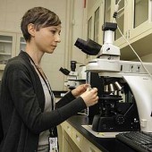
Harmony Hunter: Hi! Welcome to the podcast, I’m Harmony Hunter. It’s always fun when we get to talk about how cutting edge technology can reshape the historical narrative. So we’re excited today to introduce Kirsten Moffitt, who is the first on-staff materials analyst at the Foundation’s new Conservation Analytical Laboratory. Kirsten, thank you for being here today.
Kirsten Moffitt: It’s great to be here. Thanks for having me.
Harmony: I feel like I just said a whole mouthful of words and I’m not even sure what they mean. Tell me about the Conservation Analytical Laboratory.
Kirsten: The conservation laboratory that we just opened up is the newest lab in our conservation department. Conservation is the profession dedicated to the preservation and treatment of cultural artifacts. As you know, we have a great deal of cultural artifacts here in our collection. That can include archeological materials, architectural sites, all of the historic buildings that we have in the Historic Area, and our extensive museum collection. The analytical laboratory is a new, dedicated space that houses a suite of instrumental techniques that we can use to analyze the materials we have in our collection. I carry out analysis for the conservators, curators, and the architectural historians at the Foundation.
These projects can all vary depending on the goal of the analysis. Often the curators are interested in having me analyze an object to understand the materials that were used to make that object that can shed very helpful light on the provenance of that object: where, when, and how it was made. It can also answer some questions about the authenticity of the object or the history of the restoration of that object. Certain parts of an object can visually look the same, but chemically the can be very different, and I can actually pick that up using the instruments we that have in our lab.
A lot of the conservators come to the analytical laboratory because they need to look more closely also at the materials used to make an object they may be treating. So knowing what an object is actually made of can help them design an appropriate conservation treatment for that object to minimize the damage that could occur possibly during a treatment. It also saves them time, a lot of guesswork in trying to design that treatment.
Another reason why I might work with the conservators as well is to look at restoration materials. A lot of times, conservators need to remove previous restorations. That could include anything from perhaps a yellow varnish on a painting or an adhesive on a ceramic that’s no longer functional. They can send me very, very tiny samples from an object. These samples can actually be the size of 10 micrometers, I mean, this is the size of a speck of dust, you can barely see it. The average width of a human hair is 80 micrometers so that just gives you a sense of how small these samples are that we’re dealing with. We can learn a great deal of information from these very, very tiny, tiny samples.
The reason why these samples are so small is because when we’re working in the conservation department, we’re charged with taking care of these artifacts. We don’t ever want to remove any material that might be original. We want to avoid sampling whenever possible and actually some of the techniques that I have in the lab are completely nondestructive. If we do need to take a sample, that sample can actually be very, very small, very minimally invasive, causing little to no damage to the object.
Harmony: We’ve talked a lot about what you’re able to discover. Tell me about the tools that you’re using. They are unusual in a museum setting, or maybe they are unexpected.
Kirsten: Well, actually there are a number of museums that do have analytical laboratories, but not many. It’s really fantastic that we’ve been able to set up our own lab here at Colonial Williamsburg. We have a number of instruments that we actually use different types of energy to see how that energy interacts with the materials to learn more about our objects.
Harmony: So what’s an example of that, for somebody that’s not a scientist?
Kirsten: One of the instruments that we have in the laboratory is a handheld XRF. XRF stands for x-ray fluorescent spectroscopy. This instrument actually looks like a Star Trek ray gun. It’s about the size and shape of a hair dryer. It’s completely portable, so we can take it out of the lab. We can bring it anywhere in the Foundation to use it.
What it does is that it actually emits x-rays that can then interact with an object. It excites the elements that are within that object. The elements in the object, when they get excited, they emit their own x-rays, which are picked up by the XRF. We have a laptop that’s loaded up with analytical software. In live time, when I’m pointing that handheld XRF at, say, a silver teapot, the laptop will actually show me what’s called a spectrum. I’ll see peaks in that spectrum that correspond with the x-ray energies that are coming out of the object.
For instance, almost any element that’s on the Periodic Table that’s above Magnesium, we can identify with this technique. So if I’m looking at a silver object, I’ll see peaks on that spectrum that correspond with the energy levels for silver. I also might see little peaks for copper, because sterling silver contains certain alloys of silver and copper. Silvers that are made before the mid-19th century contain trace elements of lead, gold, and sometimes arsenic. By using this little handheld device and pointing it at a silver object, I can see peaks for silver and copper, and if I’m seeing peaks for lead, gold, and arsenic, then I can go back to the curator and say, “This spectrum that I’m seeing from this object does confirm that this object would be a colonial-era piece of silver.”
Harmony: That is fascinating.
Kirsten: Of course if those peaks are absent, that also tells us something’s going on here. The handle on this teapot may not be original to the rest of the object. Because it’s handheld, the great thing about it is we can take it out of the lab. We can bring it into the museum where objects might be too large or too delicate to be moved. We can actually point it to a painting so that we can get readings from the pigments in the paintings. So if we were to see peaks for mercury, we know that a pigment called vermillion was used. If we see peaks for copper, we know that a copper-based pigment called verdigris was used possibly. There’s really a lot of information we can get on this totally handheld device and it’s completely nondestructive. It’s a really wonderful technique for looking at our objects. We can look at glass, ceramics, just a whole wealth of objects in our collection. We can get the data very fast.
Harmony: How are these and other tools changing the way that we are able look at and understand the objects in our collection, be they metals, textiles, wood, furniture?
Kirsten: I think the fact that now this technology is actually getting to be so small and so portable, it just makes it a much more accessible technique, and much more easy to use. I just want to note that even 20 years ago, an XRF would have taken up an entire room. It was actually quite a large instrument, and you had to position the object underneath the instrument and leave the room to carry out the analysis. Now you can actually pick up the instrument and take it wherever you need to.
Another one of the instruments that we have in the lab is an FTIR microscope. That stands for “Fourier Transform Infrared Spectroscopy.” This is actually a technique that we use to irradiate the sample with an infrared light that will also generate a spectrum. The infrared light can excite certain molecular vibrations within the molecules in a sample. From that information where certain organic functional groups might vibrate, that generates little peaks on a spectrum, a spectrum similar to what we would have gotten for the XRF.
The spectrum tells us the frequencies at which these vibrations occur. That helps us understand the actual physical structure of the molecules in the material. Things like carbon hydrogen bonds or carbonate bonds, we’ll actually see peaks that correspond to these functional groups, and that helps us understand what the molecular structure of the material is. That helps us understand what it actually is. We can use this to look at organic materials like varnishes, plastics, adhesives.
Using the FTIR along with XRF, they are really wonderful complementary techniques. The XRF might be able to tell us something, for instance, contains calcium while the FTIR can tell us that it contains a carbonate. By putting those two techniques together we can understand that the material is calcium carbonate, and it’s chalk. Just having these two techniques alone in the lab can give us a really wonderful amount of information about an object. The FTIR does require a sample, but extremely, extremely small, 100-10 microns.
Another instrument that we have in the lab is a high-powered fluorescence microscope. This is a microscope that you typically see in a biological laboratory and maybe in a university. I use it for paint analysis. I carry out a lot of analysis looking at the pain layers, for instance, in our historic structures. What I can do is collect for tiny flakes of paint while I’m on site somewhere. I can essentially cut the flakes in half, set them on their sides, and look at them under this microscope to see their cross-section, essentially the sequence of layers that have been applied over time.
You’ll see the wood and the first layer of paint that would have been applied in the 18th century. Sometimes our houses have up to 25 even 30 layers of paint on them. By looking at the cross sections, we can see the layers of paint that have been applied over time. Using this high-powered microscope, I can look at not only the colors that might have originally been applied, but the distribution of pigments, the thickness of the layers, the presence or absences of varnishes.
I can also use that microscope to pull tiny fibers, from say a textile or even a paper object and examine those fibers and identify them as cotton, or wool, or silk. I can also use that microscope to look at pigments. The microscope as a magnification that goes all the way up to 1,000 times, so I can actually look at individual pigment particles and look at their physical characteristics to identify them.
I also want to note that almost all of the instruments that are in our lab were generously provided by donations from Clint and Mary Gilliland of Menlo Park, California. They provided the money that allowed us to purchase this suite of instruments. With this high-powered microscope, the FTIR, and the handheld XRF, we really have a wonderful foundation of instrumentation that we can really answer almost any question we have about the artifacts in our collection.
Harmony: It is so fascinating to hear you describe this area of conservation which I never even imagined existed.
Kirsten: A lot of people don’t.
Harmony: We’re so glad to be able to the discoveries you’re able to make in this new lab. Thank you so much for being here today.
Kirsten: Thank you.